Light Scattering Changes during Nerve Activity
& Brain Functional Optical Imaging
brief summary 1996. 11. 11
B.H.Bae
* 최근의 저의 연구 분야 입니다.
단일 뉴런에서의 광산란 현상.
다음과 같이 진폭변화와 복굴절 현상이 1968년에
처음으로 제안 되었다.
출처-1
1968년 4월 L.B. Cohen, R.D. Keynes and B.Hille, "Light Scattering and Birefringence Changes during Nerve Activity", Nature, 218,438-441,1968.
Changes in light scattering originated from at least two different phenomena, while a large part of the birefrigence change seems to be directly dependent on the potential difference across the axon membrane.
--- 복굴절의 원인으로
kerr effect등을
제안
출처-2
1968년 9월 I. Tasaki, A. Watanabe, R.Sandlin and L. Carnay, "Changes in Flurescence, Turbity, and Bireferingence associated with nerve Excitation", Proc. N.A.S, 61, 883-888, 1968.
Experimental evidence which shows that the process of excitation in the nerve is accompanied by a transient change in optical properties of the nervous tissue. The optical properties examined include flourescence, turbidity, and birefrigence.
--- 출처-1의 실험
재연
비 침습 광산란 이미징 시스템을
위한 사전조사
1. 뉴런 광
산란의 정량적 기술
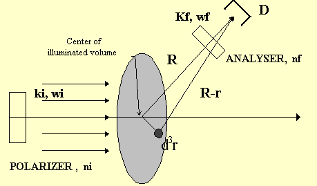
입사광 : 다음과 같이 평면파로 두자

뉴런 집단의 광학적 특성 : 유전율 텐서를 다음으로 둔다.

where,  is the dielectric
constant fluctuation tensor at position r and time t.
산란광: 긴 계산 후에 다음이 얻어진다.





Spectral Density

Scattered Light Analysis
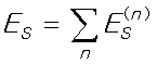
Es can be regarded as a sum of independent random variables (ES (1), ES (2), ... ).
Es ----> Gaussian distribution.
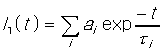
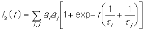
Correlation function of Scattered light
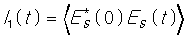
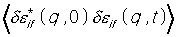

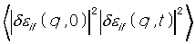
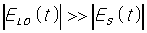
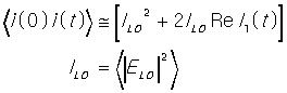
Correlation function of Scattered light + Unscattered
light

인체 조직의 광 특성
뼈를 포함한 인체는 700 nm 에서 900nm 영역이 good
window !
다음 그림은 손에 720nm의 빛을 비춘 것을 포토다이오드로
영상화 한 것. 결국 잘 투과한다는 이야기죠.
 720 nm
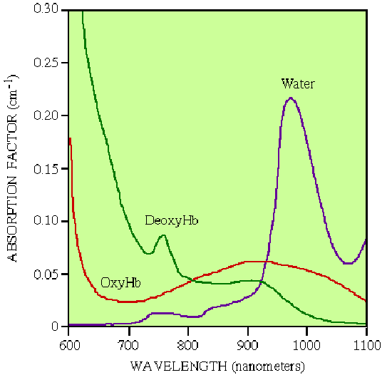
다음 그림은 인체조직에서의 물과 산소 없는 헤모글로빈과 산소가 붙어있는 헤모글로빈의 광흡수정도를 파장별로 보여주는 그림.
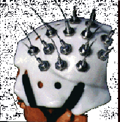
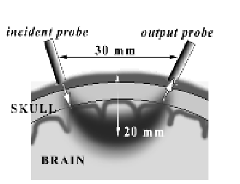
Noninvasive functional mapping with multi-channel
near infra-red spectroscopic topography in humans.
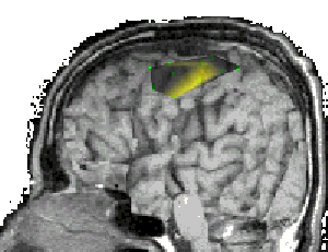
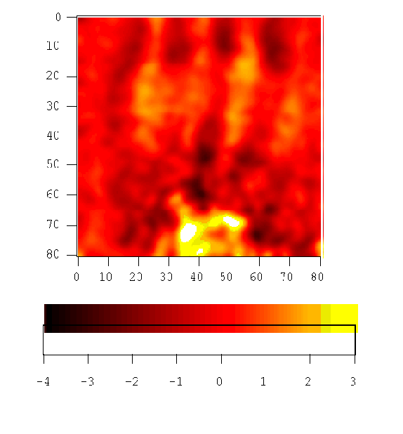
다음은 일본에서 최근에 개발된 혈액 중 헤모글로빈의 산소 소모량을 영상화 하는 장치를 보여주고 있다. 실험은 피검자가 오른쪽 손가락을 움직일때, 뇌의 어느 부위가 그 역할을 담당하는 지를 보여주고 있다. (오른쪽 그림에서 노란색으로 표현된것.)